Ratiometric in vivo auditioning of targeted silver nanoparticles
We report in the Nanoscale the in vivo application of the technology of isotopically-barcoded ratiometric silver nanoparticles for quantitative biodistribution studies. In a proof of concept study we used peptides with previously described tissue tropism; one peptide that favors vascular beds of the normal lungs (RPARPAR; receptor neuropilin-1, or NRP-1) and another that is selective for central nervous system vessels (CAGALCY). Equimolar mixtures of the peptide-targeted Ag107-NPs and Ag109 control particles were mixed and injected intravenously. Distribution profiles of Ag107 and Ag109 in tissue extracts were determined simultaneously through inductively coupled plasma mass spectrometry (ICP-MS, both on tissue extracts and on cryosections to obtain spatial information). Internally controlled ratiometric AgNP system appears to be suitable for quantitative studies of the effect of targeting ligands on NP biodistribution, at average tissue concentration and distribution at the microscopic level. The platform might be particularly relevant for target sites with high local variability in uptake, such as tumors.
Schematic representation of the concept of isotopically-barcoded silver nanoparticles for in vivo biodistribution studies (Tambet Teesalu).
Ratiometric in vivo auditioning of targeted silver nanoparticles.Toome K, Willmore AA, Paiste P, Tobi A, Sugahara KN, Kirsimäe K, Ruoslahti E, Braun GB, Teesalu T.Nanoscale. 2017 Jul 20;9(28):10094-10100. doi: 10.1039/c7nr04056c. PMID: 28695222
Hyaluronan-binding peptide for targeting peritoneal carcinomatosis
Ikemoto H, Lingasamy P, Anton Willmore AM, Hunt H, Kurm K, Tammik O, Scodeller P, Simón-Gracia L, Kotamraju VR, Lowy AM, Sugahara KN, Teesalu T.
Tumour Biol. 2017 May;39(5):1010428317701628. doi: 10.1177/1010428317701628.
Tumor-Penetrating Nanosystem Strongly Suppresses Breast Tumor Growth
Sharma S, Kotamraju VR, Mölder T, Tobi A, Teesalu T, Ruoslahti E.
Nano Lett. 2017 Mar 8;17(3):1356-1364. doi: 10.1021/acs.nanolett.6b03815. Epub 2017 Feb 17.
Linear TT1 peptide-coupled FAM-labeled iron oxide nanoparticles home to blood vessels of subcutaneous U87 tumor xenograft
Linear TT1 peptide-coupled FAM-labeled iron oxide nanoparticles (green) home to blood vessels (red) of subcutaneous U87 tumor xenograft. Cell nuclei are stained with DAPI (blue). Scale bar – 50 µm. Author – Pille Säälik PhD
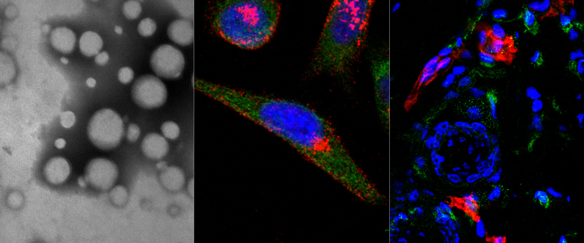
Dark-field image of silver nanoparticles with RPARPAR-MMAE-AgNP internalized by PPC-1 prostate cancer cells
Dark-field image of silver nanoparticles with cytotoxic drug monomethyl auristatin E and RPARPAR peptide (RPARPAR-MMAE-AgNP) internalized by PPC-1 prostate cancer cells after 1 h incubation. Extracellular fraction of AgNPs was removed by etching. Scale bar: 20 μm; dotted lines outline cells. Author: Allan Tobi MSc.
PET-CT image of breast tumor bearing mouse i.v. injected with iRGD targeted nanoparticles
PET-CT scan was acquired 24 hours after sample administration. The radiolabeled iRGD-nanoparticles accumulates in breast tumor (white arrow). Authors: Lorena Simón Gracia and Pablo Scodeller
FAM-UNO homes to TIE2+ tumor macrophages
Mouse model of triple negative breast cancer, 4T1. Microscope: Olympus FV1200MPE-BX61WI confocal microscope at 20X magnification Green: peptide, Red: TIE2, blue: cell nuclei. Intravenous injection. Author: Pablo Scodeller
Targeted nanosystems for imaging and therapy Spring workshop, Tartu, Estonia, May 10-12, 2017
This workshop provides an opportunity to get an overview of translational nanobiomedicine research from the leading European experts. The overarching themes are interactions of nanoparticles with biological systems, applications of precision-guided nanosystems for imaging and more efficient therapies, and the relationship of nanotechnology with personalized medicine.
Targeted nanosystems for imaging and therapy
Spring workshop, Tartu (Estonia)
May 10-12, 2017
Venue: V-Spa Hotel and Conference Center, Riia 2, Tartu
Target audience : Graduate students , research staff
Organizers: Lorena Simón Gracia This email address is being protected from spambots. You need JavaScript enabled to view it.
Tambet Teesalu This email address is being protected from spambots. You need JavaScript enabled to view it.
Free Registration at: http://registration.amarela.ee/spring-workshop/
Program and more information : Click Here
Circular structure of blood vessels homing FAM-labeled nanoparticles in P3-stem like glioblastoma
Circular structure of blood vessels homing FAM-labeled nanoparticles in P3-stem like glioblastoma. CD31 (red), FAM NPs (green), nuclei (blue). Author: Maarja Haugas, PhD
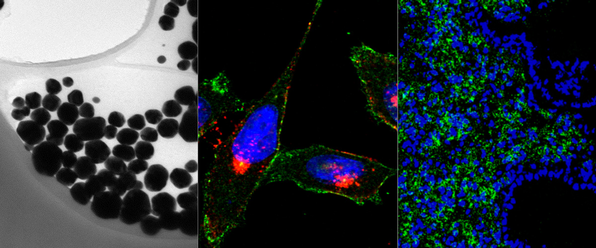
Infiltrating CD206-positive macrophages in peritoneal MKN45P gastric carcinoma xenograft
Infiltrating CD206-positive macrophages in peritoneal MKN45P gastric carcinoma xenograft.
Author: Hedi Hunt, MSc.