New p32/gC1qR Ligands for Targeted Tumor Drug Delivery
Paasonen L,Sharma S, Braun GB, Kotamraju VR, Chung TD, She ZG, Sugahara KN, Yliperttula M, Wu B, Pellecchia M, Ruoslahti E, Teesalu T
Chembiochem. 2016 Feb 19. doi: 10.1002/cbic.201500564. [Epub ahead of print]
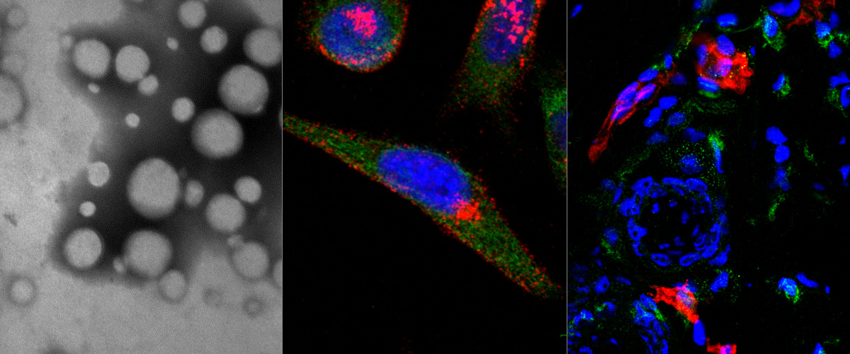
A peptide identified in the lab using in vivo phage display homes to tumor macrophages in a mouse model of a highly metastatic human gastric cancer, MKN45-P
Microscope: Olympus FV1200MPE-BX61WI confocal microscope at 60X magnification
Green: peptide, Red: macrophages, blue: cell nuclei.
Author: Pablo David Scodeller, PhD
IONW cycle of life
Microscope: SEM (magnification: 600,000x).
Author: Allan Tobi, MSc student
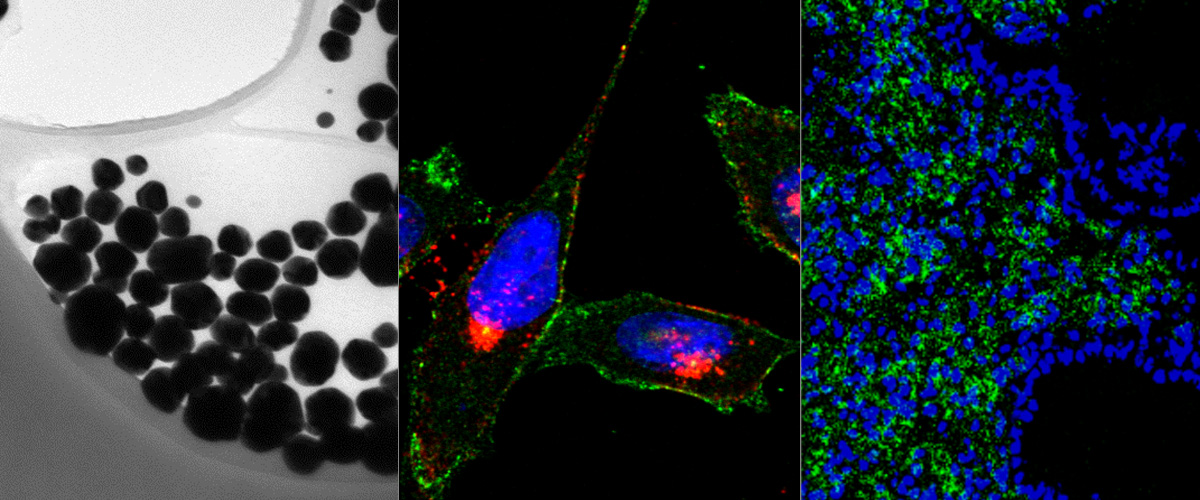
Kadri Toome's image of spheroids was shared by Molecular Probes in Facebook
Our work reaches Social Media. Few days ago Molecular Probes (Thermo Fisher Scientific) shared our image U87 cell line spheroids taking up RPARPAR-AgNPs (image of the month - April 2015 by Kadri Toome, MSc) in Facebook.
Targeted silver nanoparticles for ratiometric cell phenotyping.
Willmore AA, Simón-Gracia L, Toome K, Paiste P, Kotamraju VR, Mölder T, Sugahara KN, Ruoslahti E, Vraun GB, Teesalu T
Nanoscale. 2015 Dec 8. [Epub ahead of pront]
Expression of stem cell markers Aldh1a1 and Nestin in human stem cell like glioblastoma cell line P3
Confocal detection with Olympus FV1200MPE-BX61WI microscope and 60xoil objective.
Author: Maarja Haugas, PhD
Ex vivo visualization of targeted nanoworms homing to intraperitoneal and subcutaneous tumors after i.v. injection
Mouse model: peritoneal gastric carcinoma ( MKN-45 in Nude mice).
Author: Hedi Hunt, MSc
Multiphoton microscope at the Lab of Cancer Biology
We are excited to announce that Olympus FV1200MPE-BX61WI multiphoton microscope of the Lab of Cancer Biology is installed and fully operational. The microscope is optimized for intravital imaging of small laboratory animals and live cell imaging, including 3D imaging of fixed tissue samples. The microscope has XLPLN25xW-MP large diameter objective (NA 1.05; WD 2.00mm; Olympus) designed for multiphoton excitation, Mai Tai DeepSee Ti:Sapphire infrared laser (Spectra-Physics, tuning range 690-1040nm; pulse width 70fs), and a dedicated image analysis computer equipped with Imaris and Adobe Photoshop software. The microscope is open to all qualified researchers interested in studying dynamics of physiological and pathological processes in live multicellular organisms. Contact: Maarja Haugas (This email address is being protected from spambots. You need JavaScript enabled to view it.). Reservation and terms of use: http://www.cancerbiology.ee/tecnologies/olympus-fv1200mpe
Kadri Toome awarded the best poster prize on meeting of Finnish Peptide Society (Helsinki, August 2015)
Kadri Toome and Tambet Teesalu of the laboratory of Cancer Biology participated in the joint symposium of the Finnish Synthetic Chemistry Society, the Medicinal Chemistry Committee of the Finnish Pharmaceutical Society and the Finnish Peptide Society “Emerging targets and molecules in middle space” (Helsinki, Finland, August 24-27, 2015). Kadri’s poster “Development and in vivo validation of blood-brain barrier targeting peptides” (authors: Toome K, Säälik P, Willmore AM, Tarmo Mölder T, Sudakov A, Kõiv K, Nikonov A, Teesalu T) was awarded the best poster prize. Tambet presented an invited talk entitled „Tumor homing peptides v2.0: streamlined discovery and applications for targeted payload delivery”.
Neuropilin 1 expression (in red) in glioblastoma cell lines used in our lab
WT GBM, VEGF KO GBM and 005 – mouse-derived; P3 and NCH421K – human-derived cell lines. Blue – nuclei (DAPI).
Image acqusition: confocal system Olympus FV1200MPE.
Author: Pille Säälik, PhD