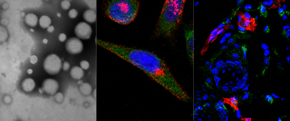
Transmission electron microscopy image of a dextran coated iron oxide nanoworm structure
Red color was applied with photo editing. Merry Christmas!
Author: Allan Tobi, MSc
Glih27 peptide conjugated with iron oxide nanoworms (green) in mouse brain with VEGF KO glioblastoma
The nanoworms with peptide home to ependymal cells. Red – blood vessels, blue – DAPI.
Author: Pille Säälik, PhD
Centre of Excellence for Genomics and Translational Medicine
University of Tartu in partnership with Estonian Biocentre and Tallinn Technical University is hosting the Centre of Excellence for Genomics and Translational Medicine
The Centre of Excellence for Genomics and Translational Medicine was awarded funding from EU European Regional Development Fund for the development of CoEs. The funding from EU is 4 818 537 EUR for the period of seven years (2016-2023).
The CoE for Genomics and Translational Medicine (GenTransMed) is based on three research groups from the Estonian Genome Center, University of Tartu and seven research groups from the Faculty of Medicine, University of Tartu. Two research groups are based in the Estonian Biocentre and the Tallinn Technical University.
The objectives of the CoE for Genomics and Translational Medicine
The CoE for Genomics and Translational Medicine stems from the need to translate discoveries in the field of genomics into improved understanding of molecular mechanisms of disease, as well as improved prevention, diagnosis and clinical care. The new interdisciplinary environment of the CoE makes it possible to interpret and rapidly utilize data produced by cutting edge omics technologies. The Centre will explore genetic and biolological mechanisms of disease by 1) investigating DNA sequence variants that contribute to disease through large scale genomic screens, and 2) using molecular and cellular bology techniques complemented by the use of animal models to study their role in disease. This offers an exciting platform for discovering important biological mechanisms that contribute to disease. The outputs of the project are anticipated to unravel processes underlying complex disease, identify new therapeutic targets and enable development of new approaches for disease prevention and precision medicine that will have a major impact in Europe and globally.
Marie Curie Actions - IRSES
7th Framework Programme for Research. technological Development and Demonstration
Project title:Binding-activated fluorescent DNA/peptide chimeric probes for cancer imaging
Co-ordinator: Universita Degli Studia Di Roma Tor Vergata Department of Chemistry Assistant Professor Dr. Francesco Ricci
Partner: University of Tartu Guest Professor Dr. Tambet Teesalu
Other Partners:
Universite de Montreal, Professor Alexis Vallēē-Bēlisle
Sanford-Burnham Medical Reserach Institute, Research Assistant Professor Dr. Kazuki Sugahara
The overriding goal of this project is to mimic Nature in the design of novel nucleic acid/peptide chimera based nanodevices that can enable cancer imaging. We will design, engineer and optimize highly specific biomolecular nanodevices that undergo binding-induced conformational changes upon target binding and, in doing so, signal the presence of the cancer marker. This scientific goal of great relevance for the clinical implications will be achieved bringing together an international and interdisciplinary group of research teams and building a collaborative environment for research, innovation and technology transfer. This program will allow the exchange of knowledge and expertise through visiting training periods for participating early-stage and experienced researchers. The training will involve each single aspect of probe development and cancer imaging. The research teams involved in this project encompass the requisite expertise and each of them has a particular focus on a single step of optical probe development and testing on cancer cells. This program will help the integration and collaboration among the research teams and the establishment of a long-term collaboration between Europe and key Third Countries. Free keywords: DNA probes, tumour recognition peptides,tumour imaging, fluorescent probes
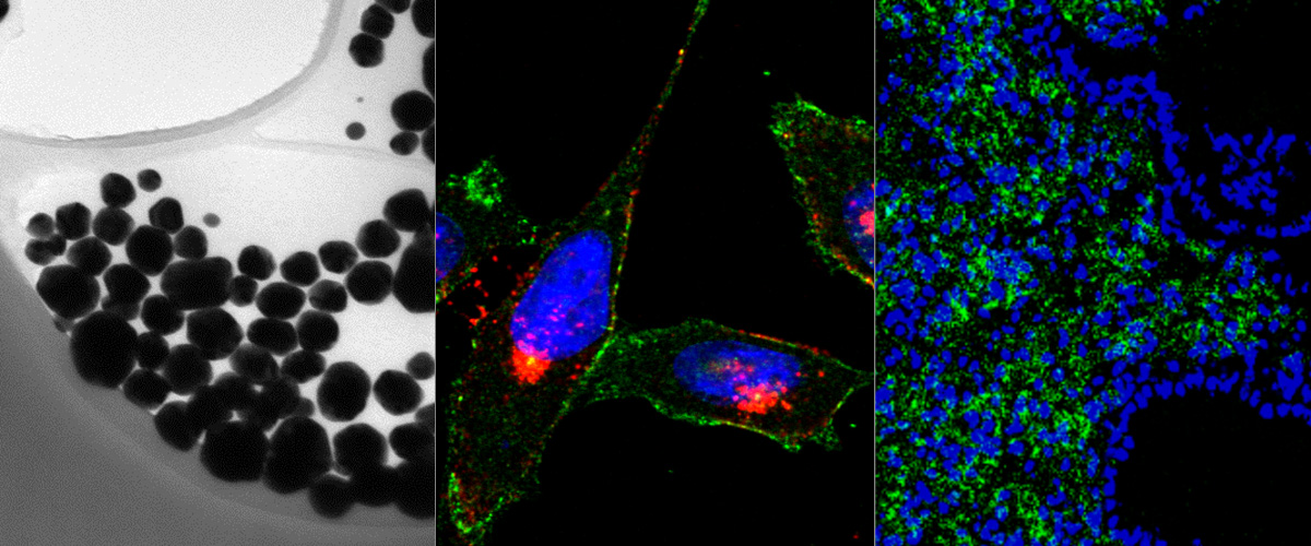
Targeting-polymeric nanoparticles are internalized in MCF-7 and U87 spheroids
Fluorescein-labeled polymeric nanoparticles functionalized with a targeting peptide (in green) are internalized into MCF-7 (from breast tumor) and U87 (from glioblastoma) spheroids that express the NRP-1 receptor (in red). The nanoparticles are observed inside the U87 cells (white arrow). Scale bar are 200 µm and 20 µm (in the big magnification).
Authors: Lorena Simón-Gracia, Ines Diaz Bessone, Kadri Toome
An infiltratively growing patient-derived mouse glioma xenograft P21 developed in our lab expresses nestin (green) and Ki67 (red) positive cells.
Cell nuclei are stained with DAPI (blue).
Author: Pille Säälik, PhD
Intravenously injected "UNO" peptide enters protumoral macrophages
Angiogenic glioblastoma, WTGBM.
Microscope: Olympus FV1200MPE-BX61WI confocal microscope at 20X magnification Green: peptide, Red: tumor associated macrophages, blue: cell nuclei.
Authors: Pablo Scodeller PhD, Pille Säälik PhD
iRGD-targeted polymersomes for enhanced intraperitoneal chemotherapy
We report in Biomaterials development of a tumor-specific delivery system for the treatment of peritoneal carcinomatosis. We demonstrate that after intraperitoneal administration, pH-sensitive polymeric vesicles loaded with an anticancer drug paclitaxel and functionalized with the tumor penetrating peptide iRGD specifically accumulate in peritoneal tumors in mice and have higher antitumor activity than free paclitaxel or Abraxane (a nano-formulation currently used in the therapy of several types of carcinoma). Our findings suggest that iRGD polymersomes may potentially be translated to therapeutic interventions against peritoneal carcinomatosis.
This collaborative study was driven and coordinated by our senior researcher Lorena Simón-Gracia and carried out together with Prof. Giuseppe Battaglia’s lab in UC London (UK) and with Drs. Kazuki N Sugahara and Ramana Kotamraju and Prof. Erkki Ruoslahti at Sanford Burnham Prebys Medical Discovery Institute in La Jolla, Calif. (USA).
iRGD peptide conjugation potentiates intraperitoneal tumor delivery of paclitaxel with polymersomes Biomaterials. 2016 Jul 20;104:247-257.
Image: Homing of green fluorescent iRGD polymersomes in CT26 peritoneal tumor (Tu). Note that control organs (liver, Li; lung, Lu; spleen, Sp; kidney, Ki) and subcutaneous tumors (Tu s.c.) show minimal labeling.
iRGD peptide conjugation potentiates intraperitoneal tumor delivery of paclitaxel with polymersomes
Simon-Gracia L, Hunt H, Scodeller P, Gaitzsch J, Kotamraju VR, Sugahara KN, Tammik O, Ruoslahti E, Battaglia G,Teesalu T
Biomaterials. 2016 Jul 20;104:247-257. doi: 10.1016/j.biomaterials.2016.07.023. [Epub ahead of print]
An homing peptide that targets injured brain tissue highlighted in Nature Reviews
This peptide can guide drugs and imaging agents to acute brain injuries and result in enhanced benefit. The peptide was identified by Aman Mann and Pablo Scodeller in the lab of Dr. Erkki Ruoslahti in collaboration with the cancer biology lab.
Link to the paper: Nature Communications
Links to news:Nature Reviews Chemical and Engineering News Novaator Science Daily The San Diego Union Tribute
Image: The tiny peptide CAQK improves the delivery of imaging agents to acute brain injuries. (Luminiscence from porous silicon nanoparticles targeted with CAQK and control peptide CGGK in brain injuries)