Second harmonic generation image of collagen fibers in mouse dermis
Detection in wavelength of 800nm with Spectra Physics MaiTai Deep See Ti:Sapphire laser. Multiphoton microscope Olympus FV1200MPE; objective XLPLN 25xW-MP (NA 1.05).
Author: Maarja Haugas, PhD
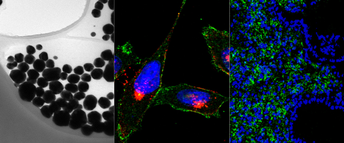
Targeted polymersomes penetrate deep into peritoneal tumors
Targeted polymersomes (in green) target Neuropilin1 rich regions (in red) in gastric peritoneal tumors when injected intraperitoneally.
Authors: Lorena Simon Gracia, PhD and Pablo Scodeller, PhD
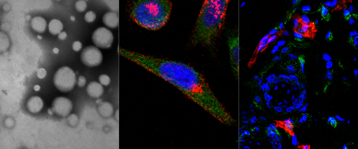
Homing of nanoworm nanoparticles to peritoneal gastric carcinoma ( MKN-45 Nude mice)
Sample collected 5h after i.p. injection.
Red: Vessels
Green: NW-TT1-FAM
Blue: Nuclei
Technology: Confocal Microscopy (Olympus FV1200MPE)
Author: Hedi Hunt, MSc
For booking please create a new event: your full name/your PI's full name/confocal or multiphoton.
The analysis computer for editing .oib/.oif files is situated in SIME building Ravila 14b-110.
1. You can log in to the computer using UT’s username and password.
2. With this computer you can access the files created with the Olympus FV1200MPE.
3. You can also access your UT’s network drives to save your files.
4. You cannot keep your edited files in the analysis computer you need to save them yourself. The files are not accessible after you have left the computer.
| Maarja Haugas, Phd: microscope information, instructions This email address is being protected from spambots. You need JavaScript enabled to view it. +372 553 5669 |
Kaarel Kurm, MSc: technical information about the microscope This email address is being protected from spambots. You need JavaScript enabled to view it. +372 515 6950 |
Kadri Toome, MSc: microscope information This email address is being protected from spambots. You need JavaScript enabled to view it. +372 5648 3748 |
For accessing the calendar please ask for invitation from: This email address is being protected from spambots. You need JavaScript enabled to view it..
The Olympus FV1200MPE multiphoton laser scanning microscope in situated in SIME building Ravila 14b-111.
- Before using the microscope please contact first with Maarja Haugas (This email address is being protected from spambots. You need JavaScript enabled to view it. ) for introductory instruction.
- To use the microscope or the analysis computer you need to book time for usage at online calendar (link below). Username and password for UT domain can be used for logging in to the microscope booking page.
- You can book the microscope maximally for 3 hours per day. In case longer time is needed, please confirm it with Maarja Haugas. If you cancel your reservation more than an hour before your booking starts, you will not be charged.
- To access the room 111, ask the key from the information secretary of SIME building.
- Please leave your outerwear in the entrance of the lab where you can also find some indoor shoes for public use. You may not enter the microscopy room with outer clothing and outdoor shoes.
- Do not enter the room with food or beverages.
- When entering the room, please make 3 steps on decontamination area (sticky matress on the floor) with both legs.
- There is a log-book next to the microscope, please fill it accordingly.
- When you have finished, please fill the log-book for how long the microscope was used and return the key. You’ll be charged only according to the time you actually used the device.
- Please clean up the place after finishing:
- When you used the 60 × oil objective, dampen the lens cleansing tissue with ethanol and wipe the objective 3 times in one direction.
- Please recycle your experiment waste yourself.
- In case of problems or errors (software, hardware), please contact Maarja Haugas or Kaarel Kurm.
There are user manuals with instructions and photos for each device (microscope, CO2 chamber, objectives). Before using the devices, please take care that you have read them through, and act accordingly.
In case you are bringing other people to the microscope room (students etc), please make sure they are familiar to the terms of using the microscope.
If you find something not in order in the room (objective not cleaned etc), please inform Maarja Haugas or Kaarel Kurm. By that you support the good laboratory practice.
There is a surveillance camera in the room.
Thanks!
| Maarja Haugas, Phd: microscope information, instructions This email address is being protected from spambots. You need JavaScript enabled to view it. +372 553 5669 |
Kaarel Kurm, MSc: technical information about the microscope This email address is being protected from spambots. You need JavaScript enabled to view it. +372 515 6950 |
Kadri Toome, MSc: microscope information This email address is being protected from spambots. You need JavaScript enabled to view it. +372 5648 3748 |
Booking: microscope can be used only during the business days from 8AM until 4PM. To book, please write your full name and your PI's full name.
For accessing the calendar please ask for an invitation from This email address is being protected from spambots. You need JavaScript enabled to view it.
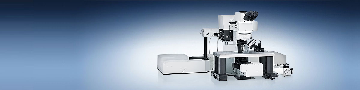
The Olympus FV1200MPE multiphoton laser scanning microscope offers brighter and clearer imaging from deep within specimens. This is thanks to optimization of the optical design for efficient multiphoton excitation and signal detection. From imaging with dedicated Olympus multiphoton objectives, to robust laser stimulation in high speed electrophysiology, uncaging and optogenetics experiments, the FV1200MPE has been optimized for scientific discovery. By closely adhering to optical principles, the Olympus FV1200MPE opens up the field of discovery science with brighter and deeper high-resolution imaging.
Olympus FV1200MPE is possible to book for imaging from Mon-Fri 8.00-16.00 BOOKING. The Microscope is placed in SIME building Ravila 14b, Tartu in room 111.
Analysis computer equiped with Imaris software is available to use from Mon-Fri 8.00-18.00 BOOKING. The analysis computer is placed in SIME building Ravila 14b, Tartu in room 110.
Usage fees:
| University of Tartu | Other | ||
| Olympus FV1200MPE: 60 minutes | 75 € | 150 €+VAT | |
| Analysis computer: 60 minutes | 7 € | 10 €+VAT | |
| Consultation and instruction: 60 minutes | 50 € | 75 €+VAT |
Contacts:
| Maarja Haugas, Phd: microscope information, instructions This email address is being protected from spambots. You need JavaScript enabled to view it. +372 553 5669 |
Kaarel Kurm, MSc: technical information about the microscope This email address is being protected from spambots. You need JavaScript enabled to view it. +372 515 6950 |
Kadri Toome, MSc: microscope information This email address is being protected from spambots. You need JavaScript enabled to view it. +372 5648 3748 |
Ultrasound molecular imaging of tumor angiogenesis with a neuropilin-1-targeted microbubble.
Zhang H, Tam S, Ingham ES, Mahakian LM, Lai CY, Tumbale SK, Teesalu T, Hubbard NE, Borowsky AD, Ferrara KW
Biomaterials. 2015 Jul;56:104-13. doi: 10.1016/j.biomaterials.2015.03.043. Epub 2015 Apr 16.
A tumor-penetrating peptide enhances circulation-independent targeting of peritoneal carcinomatosis
Sugahara KN, Scodeller P, Braun GB, de mendoza TH, Yamazaki CM, luger MD, Kitayama J, Alvarez E, Howell SB, Teesalu T, Ruoslahti E, Lowy AM
J Control Release. 2015 Jun 11;212:59-69. doi: 10.1016/j.jconrel.2015.06.009. [Epub ahead of print]
Homing of polymersome nanoparticles to peritoneal colon carcinoma (CT26 Balb/c syngeneic tumor model, sample collected 24 hours after injection)
Technology: confocal microscopy
Authors: Lorena Simon Gracia, PhD and Hedi Hunt, MSc